A new study eases fears of a link between autism and prenatal ultrasounds

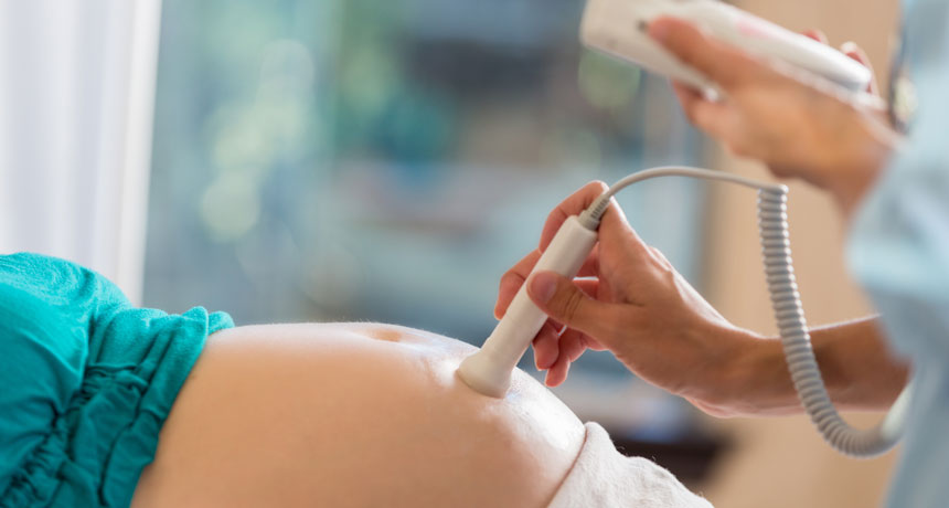
Ultrasounds during pregnancy can be lots of fun, offering peeks at the baby-to-be. But ultrasounds aren’t just a way to get Facebook fodder. They are medical procedures that involve sound waves, technology that could, in theory, affect a growing fetus.

With that concern in mind, some researchers have wondered if the rising rates of autism diagnoses could have anything to do with the increasing number of ultrasound scans that women receive during pregnancy.

The answer is no, suggests a study published online February 12 in JAMA Pediatrics. On average, children with autism were exposed to fewer ultrasounds during pregnancy, scientists found. The results should be “very reassuring” to parents, says study coauthor Jodi Abbott, a maternal fetal medicine specialist at Boston Medical Center and Boston University School of Medicine.
To back up: Autism rates have risen sharply over the last several decades (though are possibly plateauing). Against this backdrop, researchers are searching for the causes of autism — and there are probably many. Autism is known to run in families, and scientists have found some of the particular genetic hot spots that may contribute. Other factors, such as older parents and maternal obesity, can also increase the risk of autism.

Scientists suspect that in many cases, autism is caused by many factors, all working together. Could prenatal ultrasounds, which have become more routine and more powerful, be one of those factors? These scans use sound waves that penetrate mothers’ bodies, and then collect the waves that bounce back, forming a picture of fetal tissues. During this process, the waves may be able to heat up the tissue they travel through.

Work on animals has suggested that ultrasounds can in fact interfere with fetal brain development, derailing the normal movements of cells that populate the brain. Mice exposed to 30 or more minutes of ultrasound in utero had abnormal brain development, for instance. But it’s not at all clear whether a similar thing might happen in humans, and if so, whether such effects might contribute to autism.
The new study compared ultrasound exposure among three groups: 107 children diagnosed with autism spectrum disorder, 104 children diagnosed with a developmental delay, and 209 typically developing children. On average, the children with autism were exposed to 5.9 ultrasound scans over the course of pregnancy. Children with developmental delays were exposed to 6.1 scans, and typically developing children were exposed to 6.3 scans, the researchers found. (For all groups, these numbers are way above the one to two scans per low-risk pregnancy recommended by the American College of Obstetricians and Gynecologists.)

For all three groups, the duration of the scans was similar. So was the thermal index, an indication of how much warming might have happened. “In almost every parameter we looked at, ultrasound seemed perfectly safe,” says study coauthor N. Paul Rosman, a pediatric neurologist at Boston Medical Center and Boston University School of Medicine.

One measure was different, the researchers found: During the first trimester, mothers who had children with autism had slightly deeper ultrasounds than women who had typically developing children and children with developmental delays. Ultrasound depth measures the distance from the transducer paddle that emits the waves to the spot that’s being imaged. The measure “has a lot to do with the size of the mother and the distance between her skin, where the ultrasound transducer is, and where the baby is,” Abbott says.

Lots of questions remain about whether — and how — ultrasound depth, or other aspects of the technology, might affect fetuses. “The study certainly wasn’t perfect,” Rosman says. It combed back through medical records of women instead of following women from the beginning. And it didn’t control for certain traits that may influence autism, such as smoking.

The results suggest that on their own, ultrasounds don’t cause autism spectrum disorder, says Sara Jane Webb of Seattle Children’s Research Institute and the University of Washington, who cowrote a JAMA Pediatrics companion piece. “At this time, there is no evidence that ultrasound is a primary contributor to poor developmental outcomes when delivered within medical guidelines,” she says.

While there’s more science to sort out here, the news is reassuring for women who might be worried about getting scanned. Women should follow their doctors’ guidance on ultrasounds, Rosman says. “We don’t think there’s anything in this study to recommend otherwise.”

A new species of tardigrade lays eggs covered with doodads and streamers

What a spectacular Easter basket tardigrade eggs would make — at least for those celebrating in miniature.

A new species of the pudgy, eight-legged, water creatures lays pale, spherical microscopic eggs studded with domes crowned in long, trailing streamers.

Eggs of many land-based tardigrades have bumps, spines, filaments and such, presumably to help attach to a surface, says species codiscoverer Kazuharu Arakawa. The combination of a relatively plain surface on the egg itself (no pores, for instance) plus a filament crown helps distinguish this water bear as a new species, now named Macrobiotus shonaicus, he and colleagues report February 28 in PLOS ONE.
With about 20 new species added each year to the existing 1,200 or so known worldwide, tardigrades have become tiny icons of extreme survival (SN Online: 7/14/17).

“I was actually not looking for a new species,” Arakawa says. He happened on it when searching through moss he plucked from the concrete parking lot at his apartment. He routinely samples such stray spots to search for tardigrades, one of his main interests as a genome biologist at Keio University’s Institute for Advanced Biosciences in Tsuruoka City, Japan.
These particular moss-loving creatures managed to grow and reproduce in the lab —“very rare for a tardigrade,” he says. He didn’t realize it was an unknown species until he started deciphering the DNA that makes up some of its genes. The sequences he found didn’t match any in a worldwide database.

His two coauthors, at Jagiellonian University in Krakow, Poland, worked out that he had found a new member of a storied cluster of relatives of the tardigrade M. hufelandi. That species, described in 1834, kept turning up across continents around the world — or so biologists thought for more than a century. Realization eventually dawned that the single species that could live in such varied places was actually a complex of close cousins.

And now M. shonaicus adds yet another cousin to a group of about 30. Who knows where the next one will turn up. “I think there are lots more to be identified,” Arakawa says.

The debate over how long our brains keep making new nerve cells heats up

Adult mice and other rodents sprout new nerve cells in memory-related parts of their brains. People, not so much. That’s the surprising conclusion of a series of experiments on human brains of various ages first described at a meeting in November (SN: 12/9/17, p. 10). A more complete description of the finding, published online March 7 in Nature, gives heft to the controversial result, as well as ammo to researchers looking for reasons to be skeptical of the findings.

In contrast to earlier prominent studies, Shawn Sorrells of the University of California, San Francisco and his colleagues failed to find newborn nerve cells in the memory-related hippocampi of adult brains. The team looked for these cells in nonliving brain samples in two ways: molecular markers that tag dividing cells and young nerve cells, and telltale shapes of newborn cells. Using these metrics, the researchers saw signs of newborn nerve cells in fetal brains and brains from the first year of life, but they became rarer in older children. And the brains of adults had none.

There is no surefire way to spot new nerve cells, particularly in live brains; each way comes with caveats. “These findings are certain to stir up controversy,” neuroscientist Jason Snyder of the University of British Columbia writes in an accompanying commentary in the same issue of Nature.

Will Smith narrates ‘One Strange Rock,’ but astronauts are the real stars

“The strangest place in the whole universe might just be right here.” So says actor Will Smith, narrating the opening moments of a new documentary series about the wonderful unlikeliness of our own planet, Earth.

One Strange Rock, premiering March 26 on the National Geographic Channel, is itself a peculiar and unlikely creation. Executive produced by Academy Award–nominated Darren Aronofsky and by Jane Root of the production company Nutopia and narrated by Smith, the sprawling, ambitious 10-episode series is chock-full of stunningly beautiful images and CGI visuals of our dynamic planet. Each episode is united by a theme relating to Earth’s history, such as the genesis of life, the magnetic and atmospheric shields that protect the planet from solar radiation and the ways in which Earth’s denizens have shaped its surface.
The first episode, “Gasp,” ponders Earth’s atmosphere and where its oxygen comes from. In one memorable sequence, the episode takes viewers on a whirlwind journey from Ethiopia’s dusty deserts to the Amazon rainforest to phytoplankton blooms in the ocean. Dust storms from Ethiopia, Smith tells us, fertilize the rainforest. And that rainforest, in turn, feeds phytoplankton. A mighty atmospheric river, fueled by water vapor from the Amazon and heat from the sun, flows across South America until it reaches the Andes and condenses into rain. That rain erodes rock and washes nutrients into the ocean, feeding blooms of phytoplankton called diatoms. One out of every two breaths that we take comes from the photosynthesis of those diatoms, Smith adds.
As always, Smith is an appealing everyman. But the true stars of the series may be the eight astronauts, including Chris Hadfield and Nicole Stott, who appear throughout the series. In stark contrast to the colorful images of the planet, the astronauts are filmed alone, their faces half in shadow against a black background as they tell stories that loosely connect to the themes. The visual contrast emphasizes the astronauts’ roles as outsiders who have a rare perspective on the blue marble.
“Having flown in space, I feel this connection to the planet,” Stott told Science News . “I was reintroduced to the planet.” Hadfield had a similar sentiment: “It’s just one tiny place, but it’s the tiny place that is ours,” he added.
Each astronaut anchors a different episode. In “Gasp,” Hadfield describes a frightening moment during a spacewalk outside the International Space Station when his eyes watered. Without gravity, the water couldn’t form into teardrops, so it effectively blinded him. To remove the water, he was forced to allow some precious air to escape his suit. It’s a tense moment that underscores the pricelessness of the thin blue line, visible from space, that marks Earth’s atmosphere. “It contains everything that’s important to us,” Hadfield says in the episode. “It contains life.”

Stott, meanwhile, figures prominently in an episode called “Storm.” Instead of a weather system, the title refers to the rain of space debris that Earth has endured throughout much of its history — including the powerful collision that formed the moon (SN: 4/15/17, p. 18). Stott describes her own sense of wonder as a child, watching astronauts land on our closest neighbor — and how the travels of those astronauts and the rocks they brought back revealed that Earth and the moon probably originated from the same place.

It’s glimpses like these into the astronauts’ lives and personalities — scenes of Hadfield strumming “Space Oddity” on a guitar, for example, or Stott chatting with her son in the family kitchen — that make the episodes more than a series of beautiful and educational IMAX films. Having been away from the planet for a short time, the astronauts see Earth as precious, and they convey their affection for it well. Stott said she hopes that this will be the ultimate takeaway for viewers, for whom the series may serve as a reintroduction to the planet they thought they knew so well. “I hope that people will … appreciate and acknowledge the significance of [this reintroduction],” she said, “that it will result in an awareness and obligation to take care of each other.”
Editor’s note: This story was updated on March 19, 2018, to add a mention of a second executive producer.

Venus may be home to a new kind of tectonics

THE WOODLANDS, Texas — Venus’ crust is broken up into chunks that shuffle, jostle and rotate on a global scale, researchers reported in two talks March 20 at the Lunar and Planetary Science Conference.

New maps of the rocky planet’s surface, based on images taken in the 1990s by NASA’s Magellan spacecraft, show that Venus’ low-lying plains are surrounded by a complex network of ridges and faults. Similar features on Earth correspond to tectonic plates crunching together, sometimes creating mountain ranges, or pulling apart. Even more intriguing, the edges of the Venusian plains show signs of rubbing against each other, also suggesting these blocks of crust have moved, the researchers say.
“This is a new way of looking at the surface of Venus,” says planetary geologist Paul Byrne of North Carolina State University in Raleigh.

Geologists generally thought rocky planets could have only two forms of crust: a stagnant lid as on the moon or Mars — where the whole crust is one continuous piece — or a planet with plate tectonics as on Earth, where the surface is split into giant moving blocks that sink beneath or collide with each other. Venus was thought to have one solid lid (SN: 12/3/11, p. 26).

Instead, those options may be two ends of a spectrum. “Venus may be somewhere in between,” Byrne said. “It’s not plate tectonics, but it ain’t not plate tectonics.”

While Earth’s plates move independently like icebergs, Venus’ blocks jangle together like chaotic sea ice, said planetary scientist Richard Ghail of Imperial College London in a supporting talk.
Ghail showed similar ridges and faults around two specific regions on Venus that resemble continental interiors on Earth, such as the Tarim and Sichuan basins in China. He named the two Venusian plains the Nuwa Campus and Lada Campus. (The Latin word campus translates as a field or plain, especially one bound by a fence, so he thought it was fitting.)
Crustal motion may be possible on Venus because the surface is scorching hot (SN: 3/3/18, p. 14). “Those rocks already have to be kind of gooey” from the high temperatures, Byrne said. That means it wouldn’t take a lot of force to move them. Venus’ interior is also probably still hot, like Earth’s, so convection in the mantle could help push the blocks around.

“It’s a bit of a paradigm shift,” says planetary scientist Lori Glaze of NASA’s Goddard Space Flight Center, who was not involved in the new work. “People have always wanted Venus to be active. We believe it to be active, but being able to identify these features gives us more of a sense that it is.”

The work may have implications for astronomers trying to figure out which Earth-sized planets in other solar systems are habitable (SN: 4/30/16, p. 36). Venus is almost the same size and mass as the Earth. But no known life exists on Venus, where the average surface temperature is 462° Celsius and the atmosphere is acidic. Scientists have long speculated that the planet’s apparent lack of plate tectonics might play a role in making the planet so seemingly uninhabitable.

What’s more, the work also underlines the possibility that planets go through phases of plate tectonics (SN: 6/25/16, p. 8). Venus could have had plate tectonics like Earth 1 billion or 2 billion years ago, according to a simulation presented at the meeting by geophysicist Matthew Weller of the University of Texas at Austin.

“As Venus goes, does that predict where the Earth is going in the relatively near future?” he wondered.

A single atom can gauge teensy electromagnetic forces

Zeptonewton
ZEP-toe-new-ton n.
A unit of force equal to one billionth of a trillionth of a newton.

An itty-bitty object can be used to suss out teeny-weeny forces.

Scientists used an atom of the element ytterbium to sense an electromagnetic force smaller than 100 zeptonewtons, researchers report online March 23 in Science Advances. That’s less than 0.0000000000000000001 newtons — with, count ‘em, 18 zeroes after the decimal. At about the same strength as the gravitational pull between a person in Dallas and another in Washington, D.C., that’s downright feeble.
After removing one of the atom’s electrons, researchers trapped the atom using electric fields and cooled it to less than a thousandth of a degree above absolute zero (–273.15° Celsius) by hitting it with laser light. That light, counterintuitively, can cause an atom to chill out. The laser also makes the atom glow, and scientists focused that light into an image with a miniature Fresnel lens, a segmented lens like those used to focus lighthouse beams.

Monitoring the motion of the atom’s image allowed the researchers to study how the atom responded to electric fields, and to measure the minuscule force caused by particles of light scattering off the atom, a measly 95 zeptonewtons.

Why cracking your knuckles can be so noisy

“Pop” goes the knuckle — but why?

Scientists disagree over why cracking your knuckles makes noise. Now, a new mathematical explanation suggests the sound results from the partial collapse of tiny gas bubbles in the joints’ fluid.

Most explanations of knuckle noise involve bubbles, which form under the low pressures induced by finger manipulations that separate the joint. While some studies pinpoint a bubble’s implosion as the sound’s source, a paper in 2015 showed that the bubbles don’t fully implode. Instead, they persist in the joints up to 20 minutes after cracking, suggesting it’s not the bubble’s collapse that creates noise, but its formation (SN: 5/16/15, p. 16).
But it wasn’t clear how a bubble’s debut could make sounds that are audible across a room. So two engineers from Stanford University and École Polytechnique in Palaiseau, France, took another crack at solving the mystery.

The sound may come from bubbles that collapse only partway, the two researchers report March 29 in Scientific Reports. A mathematical simulation of a partial bubble collapse explained both the dominant frequency of the sound and its volume. That finding would also explain why bubbles have been observed sticking around in the fluid.

Comb jellies have a bizarre nervous system unlike any other animal

Shimmering, gelatinous comb jellies wouldn’t appear to have much to hide. But their mostly see-through bodies cloak a nervous system unlike that of any other known animal, researchers report in the April 21 Science.

In the nervous systems of everything from anemones to aardvarks, electrical impulses pass between nerve cells, allowing for signals to move from one cell to the next. But the ctenophores’ cobweb of neurons, called a nerve net, is missing these distinct connection spots, or synapses. Instead, the nerve net is fused together, with long, stringy neurons sharing a cell membrane, a new 3-D map of its structure shows.
While the nerve net has been described before, no one had generated a high-resolution, detailed picture of it.

It’s possible the bizarre tissue represents a second, independent evolutionary origin of a nervous system, say Pawel Burkhardt, a comparative neurobiologist at the University of Bergen in Norway, and colleagues.

Superficially similar to jellyfish, ctenophores are often called comb jellies because they swim using rows of beating, hairlike combs. The enigmatic phylum is considered one of the earliest to branch off the animal tree of life. So ctenophores’ possession of a simple nervous system has been of particular interest to scientists interested in how such systems evolved.

Previous genetics research had hinted at the strangeness of the ctenophore nervous system. For instance, a 2018 study couldn’t find a cell type in ctenophores with a genetic signature that corresponded to recognizable neurons, Burkhardt says.

Burkhardt, along with neurobiologist Maike Kittelmann of Oxford Brookes University in England and colleagues, examined young sea walnuts (Mnemiopsis leidyi) using electron microscopes, compiling many images to reconstruct the entire net structure. Their 3-D map of a 1-day-old sea walnut revealed the funky synapse-free fusion between the five sprawling neurons that made up the tiny ctenophore’s net.
The conventional view is that neurons and the rest of the nervous system evolved once in animal evolutionary history. But given this “unique architecture” and ctenophores’ ancient position in the animal kingdom, it raises the possibility that nerve cells actually evolved twice, Burkhardt says. “I think that’s exciting.”

But he adds that further work — especially on the development of these neurons — is needed to help verify their evolutionary origin.

The origins of the animal nervous system is a murky area of research. Sponges — the traditional competitors for the title of most ancient animal — don’t have a nervous system, or muscles or fundamental vision proteins called opsins, for that matter. But there’s been mounting evidence to suggest that ctenophores are actually the most ancient animal group, older even than sponges (SN: 12/12/13).

If ctenophores arose first, it “implies that either sponges have lost a massive number of features, or that the ctenophores effectively evolved them all independently,” says Graham Budd, a paleobiologist at Uppsala University in Sweden who was not involved in the research.

If sponges emerged first, it’s still possible that ctenophores evolved their nerve net independently rather than inheriting it from a neuron-bearing ancestor, Burkhardt says. Ctenophores have other neurons outside the nerve net, such as mesogleal neurons embedded in a ctenophore’s gelatinous body layer and sensory cells, the latter of which may communicate with the nerve net to adjust the beating of the combs. So, it’s possible they’re a mosaic of two nervous systems of differing evolutionary origins.

But Joseph Ryan, a bioinformatician at the University of Florida in Gainesville, doesn’t think the results necessarily point to the parallel evolution of a nervous system. Given how long ctenophores have been around — especially if they are older than sponges — the ancestral nervous system may have had plenty of time to evolve into something weird and highly-specialized, says Ryan, who was not part of the study. “We’re dealing with close to a billion years of evolution. We’re going to expect strange things to happen.”

The findings are “one more bit of the jigsaw puzzle,” Budd says. “There’s a whole bunch we don’t know about these rather common and rather well-known animals.”

For instance, it’s unclear how the nerve net works. Our neurons use rapid changes in voltage across their cell membranes to send signals, but the nerve net might work quite differently, Burkhardt says.

There are reports of potentially similar systems in other animals, such as by-the-wind-sailor jellies (Velella velella). Studying them in detail, along with nerve nets in other ctenophore species, could determine just how unusual this synapse-less nervous system is.

Northern elephant seals sleep just two hours a day at sea

Northern elephant seals are the true masters of the power nap.

On long trips out to sea, the seals snooze less than 20 minutes at a time, researchers report in the April 21 Science. The animals average just two hours of shut-eye per day while swimming offshore for months — rivaling African elephants for the least sleep measured among mammals (SN: 3/1/17).

“It’s important to map these extremes of [sleep behavior] across the animal kingdom to get a better sense of the evolution and the function of sleep for all mammals, including humans,” says Jessica Kendall-Bar, an ecophysiologist at the University of California, San Diego. Knowing how seals catch their z’s could also guide conservation efforts to protect places where they sleep.
Northern elephant seals (Mirounga angustirostris) spend most of the year out in the Pacific Ocean. On these odysseys, the animals forage around the clock for fish, squid and other food to sustain their enormous bodies, which can be as hefty as a car (SN: 2/4/22). Because northern elephant seals are most vulnerable to sharks and killer whales at the surface, they come up for air only a couple minutes at a time between 10- to 30-minute deep dives (SN: 9/28/02).

“People had known that these seals dive almost all the time when they’re out in the ocean, but it wasn’t known if and how they sleep,” says Niels Rattenborg, a neurobiologist at the Max Planck Institute for Biological Intelligence in Seewiesen, Germany, who was not involved in the study.

To find out if the seals sleep while diving, Kendall-Bar and her colleagues developed a watertight EEG cap for the animals. Using the cap and other sensors, the team tracked the brain waves, heart rates and 3-D motion of 13 young female seals, including five at a lab and six hanging out at coastal Año Nuevo State Park north of Santa Cruz, Calif. EEG data recorded while seals were slumbering revealed what the animals’ naptime brain waves looked like.

Kendall-Bar’s team also took two sensor-strapped seals from Año Nuevo and released them at another beach about 60 kilometers south. To swim home, the seals had to cross the deep Monterey Canyon — a locale similar to the deep, predator-fraught waters frequented by seals on months-long foraging trips. Matching the seals’ EEG readings to their diving motions on this journey showed how northern elephant seals sleep on long voyages.

The animals first swim 60 to 100 meters below the surface, then relax into a glide, Kendall-Bar says. As they nod off into slow-wave sleep, the animals keep holding themselves upright for several minutes. But as REM sleep sets in, so does sleep paralysis. The animals flip upside-down and drift in gentle spirals toward the seafloor. Seals can descend hundreds of meters deep during these naps — far below where their predators normally prowl. When the seals wake after five to 10 minutes of sleep, they swim up to the surface. The whole routine takes about 20 minutes.

Looking for that distinct sleep dive motion, the researchers could pick out naps in the dive records of 334 adult seals that had been outfitted with tracking tags from 2004 to 2019. Those sleep patterns revealed that northern elephant seals conk out, on average, around two hours per day while on months-long foraging missions. But the seals sleep nearly 11 hours per day while on land to mate and molt, where they can indulge in long, beachside siestas without worrying about predators.
“What the seals are doing might be something like what we do when we sleep in on the weekend, but it’s on a much longer timescale,” Rattenborg says. He and his colleagues have found a similar feast-and-famine style of sleep in great frigate birds, which fly over the ocean (SN: 6/30/16). “Although they can sleep while they’re flying,” he says, “they sleep less than an hour a day for up to a week at a time, and once back on land, they sleep over 12 hours a day.”

Curiously, northern elephant seals’ sleep habits are quite different from how other marine mammals have been seen sleeping in labs. “Many of them … sleep in just half of their brain at a time,” Kendall-Bar says. That half-awake state allows dolphins, fur seals and sea lions to practice constant vigilance, literally sleeping with one eye open.

“I think it’s pretty cool that elephant seals are doing this without [one-sided] sleep,” Kendall-Bar says. “They’re shutting off both halves of their brain completely and leaving themselves vulnerable.” It seems the key to enjoying such deep sleep is sleeping deep in the sea.

Cosmic antimatter hints at origins of huge bubbles in our galaxy’s center

MINNEAPOLIS — Bubbles of radiation billowing from the galactic center may have started as a stream of electrons and their antimatter counterparts, positrons, new observations suggest. An excess of positrons zipping past Earth suggests that the bubbles are the result of a burp from our galaxy’s supermassive black hole after a meal millions of years ago.

For over a decade, scientists have known about bubbles of gas, or Fermi bubbles, extending above and below the Milky Way’s center (SN: 11/9/10). Other observations have since spotted the bubbles in microwave radiation and X-rays (SN: 12/9/20). But astronomers still aren’t quite sure how they formed.
A jet of high-energy electrons and positrons, emitted by the supermassive black hole in one big burst, could explain the bubbles’ multi-wavelength light, physicist Ilias Cholis reported April 18 at the American Physical Society meeting.

In the initial burst, most of the particles would have been launched along jets aimed perpendicular to the galaxy’s disk. As the particles interacted with other galactic matter, they would lose energy and cause the emission of different wavelengths of light.

Those jets would have been aimed away from Earth, so those particles can never be detected. But some of the particles could have escaped along the galactic disk, perpendicular to the bubbles, and end up passing Earth. “It could be that just now, some of those positrons are hitting us,” says Cholis, of Oakland University in Rochester, Mich.

So Cholis and Iason Krommydas of Rice University in Houston analyzed positrons detected by the Alpha Magnetic Spectrometer on the International Space Station. The pair found an excess of positrons whose present-day energies could correspond to a burst of activity from the galactic center between 3 million and 10 million years ago, right around when the Fermi bubbles are thought to have formed, Cholis said at the meeting.

The result, Cholis said, supports the idea that the Fermi bubbles came from a time when the galaxy’s central black hole was busier than it is today.