Most people think that autism is a disorder of the brain. But the skin may play a role, too, a new study suggests.
Nerve cells in the skin are abnormal in mice with mutations in autism-related genes, leading to poor touch perception, scientists report June 9 in Cell. This trouble sensing touch may influence the developing brain in a way that leads to social deficits and anxiety later in life.
The results raise the provocative idea that fixing abnormal senses may alleviate some of the behavioral symptoms of autism, says study coauthor David Ginty, a neuroscientist at Harvard Medical School. To explore the role of touch, Ginty and colleagues used mice that carried mutations in genes linked to autism. The genes are active in many places, including the brain. But the researchers used genetic tricks to place the mutated genes only in the peripheral nervous system — the collections of nerves outside the brain and spinal cord.
Adding mutations in a handful of autism-related genes only in peripheral nerves interfered with the mice’s sense of touch. These mice had trouble telling a smooth object from a rough one, and they had outsized reactions to harmless puffs of air. “They’re really touchy when you pick them up,” Ginty says. The sensory breakdown was caused by touch-sensing nerve cells that seemed to have trouble sending messages to the spinal cord, the researchers found.
Some mice also had behavioral deficits. Those with mutations in one of two genes — Mecp2 or Gabrb3 — in the peripheral nervous system, but not the brain, showed more signs of anxiety and interacted with other mice less than mice that didn’t have those mutations. Discovering that changes in the touch-sensing nerve cells could affect behavior was unexpected, Ginty says.
The skin’s influence seems to be important early in life. Social behaviors and anxiety didn’t suffer when the genes were first mutated in touch-sensing nerve cells during adulthood. The effect on behavior showed up only when the genes were abnormal during development, the team found.
That finding is “the most impressive part of the work,” says neuroscientist Kevin Pelphrey of George Washington University in Washington, D.C. The results emphasize how autism is an inherently developmental disorder, he says. Pelphrey and colleagues previously found that the brains of children with autism react differently to light touch, which fits with the idea that problems of touch may be involved in the disorder.
Next, Ginty and colleagues plan to figure out exactly when these genes do their important work in the peripheral nervous system. “We are now really interested in the window of time,” he says. “Presumably that window closes at some point, and we’re trying to figure out when that is.” The researchers will also explore ways to restore normal touch sensation, including drugs or genetic manipulations, that would work before the window closes.
It’s possible that other nerve cells outside the brain are affected in autism, too, says neuroscientist Aaron McGee of the University of Southern California in Los Angeles. “If you have these problems with peripheral nerves that have roles in active sensation, do you also have problems with the nerves that innervate the gut?” If so, that could help explain why people with autism often experience gut trouble.
McGee cautions that it’s difficult to compare behaviors of mice with symptoms of autism in people. But he says that the genetic experiments described in the paper are “awesome, thorough and significant.”
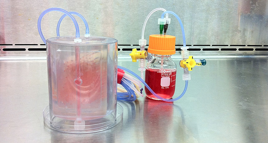
Pig fat has made the leap from kitchen staple to laboratory marvel for its ability to grow bone. Stem cells from the fat tissue of Yucatán minipigs grew into pieces of bone that were then successfully implanted into the pigs’ jaws, researchers report June 15 in Science Translational Medicine.
The team of bioengineers used cow bone scaffolds infused with stem cells from a minipig’s fat tissue to grow bone grafts in a special chamber in the lab. The new bones, which were personally fitted to each minipig’s jaw, fared better after six months than standard bone grafts not seeded with fat cells.
The new research brings scientists a step closer to one day using fat stem cells to repair humans’ broken or worn-out body parts.
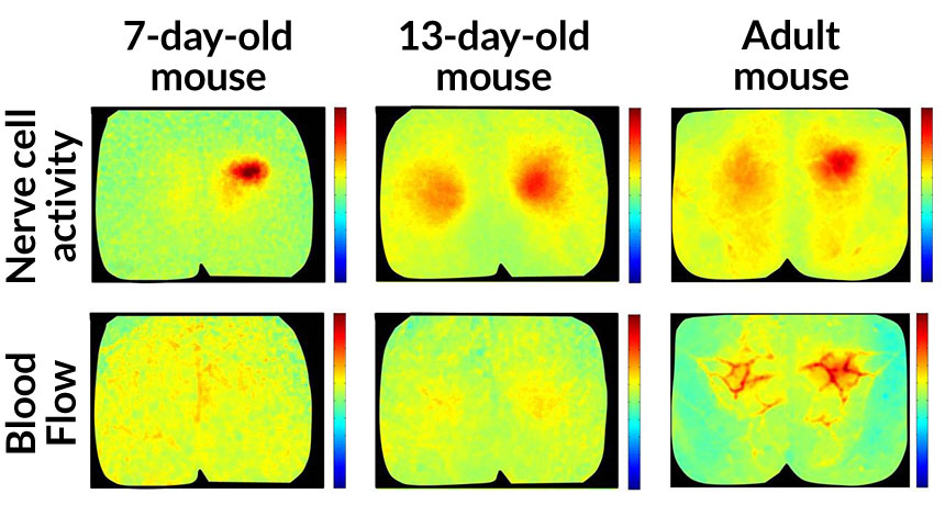
Busy nerve cells in the brain are hungry and beckon oxygen-rich blood to replenish themselves. But active nerve cells in newborn mouse brains can’t yet make this request, and their silence leaves them hungry, scientists report June 22 in the Journal of Neuroscience.
Instead of being a dismal starvation diet, this lean time may actually spur the brain to develop properly. The new results, though, muddy the interpretation of the brain imaging technique called functional MRI when it is used on infants. Most people assume that all busy nerve cells, or neurons, signal nearby blood vessels to replenish themselves. But there were hints from fMRI studies of young children that their brains don’t always follow this rule. “The newborn brain is doing something weird,” says study coauthor Elizabeth Hillman of Columbia University.
That weirdness, she suspected, might be explained by an immature communication system in young brains. To find out, she and her colleagues looked for neuron-blood connections in mice as they grew. “What we’re trying to do is create a road map for what we think you actually should see,” Hillman says.
When 7-day-old mice were touched on their hind paws, a small group of neurons in the brain responded instantly, firing off messages in a flurry of activity. Despite this action, no fresh blood arrived, the team found. By 13 days, the nerve cell reaction got bigger, spreading across a wider stretch of the brain. Still the blood didn’t come. But by the time the mice reached adulthood, neural activity prompted an influx of blood. The results show that young mouse brains lack the ability to send blood to busy neurons, a skill that influences how the brain operates (SN: 11/14/15, p. 22).
That finding was enabled by technology that allowed the researchers to see neural activity and blood flow at the exact same time. It’s “a powerful application of cutting-edge imaging techniques,” says neuroscientist Alan Jasanoff of MIT.
Showing that oxygen demands are unheeded during early development is interesting, says neuroscientist Matthew Colonnese of George Washington University School of Medicine and Health Sciences in Washington, D.C. More studies are needed to say whether human infant brains behave similarly and, if so, how this process might sculpt the brain.
The results don’t mean that fMRI data from young children aren’t valuable, Hillman says. “What we are begging people to do is to make room for this hypothesis, and actually treat it as an opportunity.” Blood flow data might not be a good proxy for neural activity in newborns, but “it may well be measuring a change that is very important to normal brain development,” she says.
Some insects make dirt look like — well, dirt. And they’ve been doing it for a while.
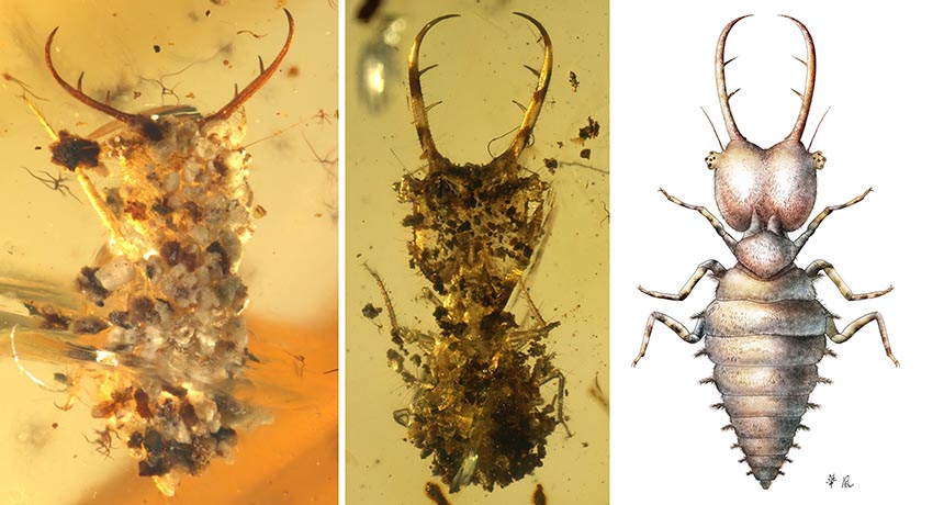
Donning a bit of debris to blend in with the environment is common practice for a subset of insects and other creepy-crawlies trying to hide from predators. (Crabs, spiders and snails do it, too.) To investigate when this behavior originated, Bo Wang of the Chinese Academy of Sciences and colleagues examined insects preserved in amber from Burma, France and Lebanon that date back 100 million years to the Cretaceous period.
Out of 300,000 insect specimens examined, 39 of them sported what appear to be dirt and vegetation disguises. Anatomical analysis suggests that these insects are early relatives of lacewings, assassin bugs and owlflies. The ancient critters decorated themselves with soil, sand, bits of wood and even tiny ferns, the team reports June 24 in Science Advances.
Until now, only one preserved, dirt-decorated insect from the Mesozoic era had been discovered. But the new finds suggest that this behavior was already widespread in some insect families back then.
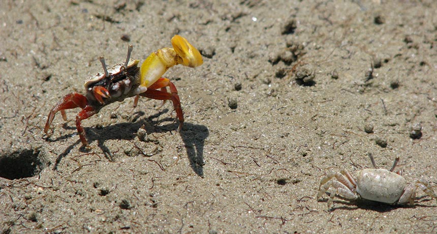
Among people, a man stepping aside to let a woman pass through a door first is seen as a gentlemanly — if a bit old-fashioned — act. Among banana fiddler crabs, though, this behavior is a trap — one that lets a male crab coerce a female into a mating she may not have preferred.
To catch the attention of a female and lure her into his burrow, a male banana fiddler crab stands outside the entrance to his cave and waves the larger of his two claws. A female will look him over and consider his size, the color of his claw and how he’s waving it. If she likes what she sees, she’ll approach him. She might decide to enter his burrow and check it out, and once inside, she might stick around for mating if she thinks that the burrow has the right conditions for rearing her embryos.
When a female approaches a male and his burrow, most males enter first, letting their potential mate follow him down. But many male crabs take another approach, stepping aside and following her into the lair — letting a male trap the female inside and mate with her, researchers report June 15 in PLOS ONE.
Christina Painting of the Australian National University in Canberra and colleagues observed banana fiddler crabs in Darwin, Australia, during two mating seasons, watching what happened as males waved their claws and females made their choice. When a female was interested in a male, the guys entered the burrow first 32 percent of the time. While females were more likely to enter a burrow if a male entered first (71 percent versus only 41 percent when the guy stepped aside), the trapping strategy was more successful in getting a mating out of the meeting. When the male followed the female in, 79 percent of females stuck around the mate. But waiting for her to follow resulted in a pairing only 54 percent of the time.
“The results strongly suggest that entering a male’s burrow first reduces the probability that a female will leave the burrow after sampling it since females are effectively trapped underground in the narrow burrow shaft when the male follows her in,” the researchers write.
So why would a female ever enter a burrow first if there were the possibility that she would be trapped inside and coerced into mating? Perhaps this might give the female a chance to test the male’s strength, the researchers suggest. If she can successfully fight her way out, then the male was obviously not worthy of her attention. Or it is possible that this is just a type of courtship behavior in which no coercion is actually happening. It’s difficult to know exactly what’s going on underground.
This isn’t the first time that the males of a fiddler crab species have been found behaving in what we might consider an ungentlemanly fashion. Males of other species have been found trapping, herding, startling and capturing females in their attempts to coerce a mating. And some male sand bubbler crabs, the researchers note, have even been found behaving somewhat like pirates of the sand-mud flats: Males have been spotted capturing female crabs, carrying them back to their burrows and forcing them into their underground lairs for mating.
Olympic swimmers shave their bodies before a big race to break records. Swordfish use a different trick, a new study suggests: They grease their heads. The fish (Xiphias gladius) are among the fastest in the ocean — their streamlined bodies can cut through the water at about 90 kilometers per hour.
A newly discovered oil-producing organ in the fish’s head gives it slick skin that could boost its speed, scientists report in the July 6 Journal of Experimental Biology. MRI scans show that the organ links to tiny pores on the head that ooze the oil, creating a thin layer of lubrication on the skin’s surface. Tiny ridged structures called denticles surround the pores. Denticles look like scales but are made of dentine and enamel, like teeth. The scientists, a team from the Netherlands, think the lubrication and the textured denticles might work together, making a water-repelling surface that lets swordfish glide through the water with minimal drag.
If you’ve ever watched a baby purse her lips to hoot for the first time, or flash a big, gummy grin when she sees you, or surprise herself by rolling over, you’ve glimpsed the developing brain in action. A baby’s brain constructs itself into something that controls the body, learns and connects socially.
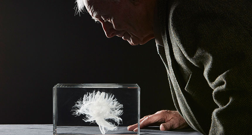
Spending time with an older person, you may notice signs of slippage. An elderly man might forget why he went into the kitchen, or fail to anticipate the cyclist crossing the road, or muddle medications with awkward and unfamiliar names. These are the signs of the gentle yet unrelenting neural erosion that comes with normal aging. These two seemingly distinct processes — development and aging — may actually be linked. Hidden in the brain-building process, some scientists now suspect, are the blueprints for the brain’s demise. The way the brain is built, recent research suggests, informs how it will decline in old age. That the end can be traced to the beginning sounds absurd: A sturdily constructed brain stays strong for decades. During childhood, neural pathways make connections in a carefully choreographed order. But in old age, this sequence plays in reverse, brain scans reveal. In both appearance and behavior, old brains seem to drift backward toward earlier stages of development. What’s more, some of the same cellular tools are involved in both processes.
Probing the connections between growing and aging may reveal how time affects the brain. And with a deeper understanding of brain aging, and the tools involved, scientists might be able to slow — or even stop — mental decline. That’s a lofty goal, made even more challenging by the multitude of theories from a diversity of researchers that aim to explain why and how the brain ages. Everybody focuses on a different aspect of the aging brain, leaving no one with a sense of the whole process, says epigeneticist Art Petronis of the Center for Addiction and Mental Health in Toronto. It’s like people trying to put together a giant jigsaw puzzle from separate rooms, each with only a few pieces in hand. So far, people studying how the brain ages have found only the evidence they can grab.
Petronis and others are intrigued by the idea that the brain’s early life holds clues to its end. “You see blips here and blips there,” he says. “This critical mass is accumulating.”
Other scientists, including Caleb Finch of the University of Southern California, in Los Angeles, caution against falling for appealing but overly simple explanations for aging. As a gerontologist who has been thinking about aging for 50 years, he has seen aging theories come and go, a perspective that makes him skeptical that the complex process can be reduced to the notion that it’s just development in reverse. “The more we poke into biology, the more wondrously complex it is,” he says.
Nonetheless, there’s something to the notion that aging starts early. “We are born dying,” Finch says. And poking at that idea just might lead somewhere.
Head start When the human brain makes its first appearance in the third week of gestation, it is no more than a minuscule smear of indistinct cells. This glob then grows at a furious rate up through the preschool years. At the same time, these accumulating brain cells begin to take on specific jobs, changing from generalists to specialists. Nerve cells are born and migrate to their final destinations, linking up in precise order to form the high-speed neural connections that enable memory, emotion and thought. And scientists now realize that the way the brain is built has lifelong effects. In 1932 and 1947, nearly every Scottish 11-year-old sat down to take an intelligence test. Decades later, their scores have matured into academic gold, offering scientists a rare opportunity to see how intelligence fares with age. In 1999, scientists led by Ian Deary of the University of Edinburgh got back in touch with as many of the long-ago test takers as possible, forming a group of more than 1,000 people — ranging in age from 80 to 95 — called the Lothian Birth Cohort. Deary and colleagues have studied the group in detail, and one factor rises above the rest: People with higher intelligence scores at age 11 were more likely to have better thinking skills in old age.
Childhood intelligence wasn’t the only factor, though. From the start, Deary and his colleagues cast a wide net, imaging participants’ brains and examining genetics, lifestyles, health and social factors. “We were right to do so, because there is a large range of mostly small influences on people’s cognitive aging,” he says. But the fact that intelligence at age 11 can partially predict who will be sharp into their 90s suggests that a long-lasting brain must be solidly constructed.
One way in which the brain is built well involves its white matter — tracts of tissue that connect distant brain regions, allowing for quick communication. And in fact, members of the Lothian Birth Cohort with healthier white matter in old age, measured by an MRI-based brain scan method called diffusion tensor imaging, performed better on tests of brain function, Deary and colleagues found.
Mature neural highways take decades to develop. Brain areas are still solidifying into a person’s thirties. The later-blooming brain regions oversee jobs like impulse control and judgment, two well-known weak spots among teenagers.
These slow-to-grow brain networks are the first to go in old age, neuroscientist Gwenaëlle Douaud of the University of Oxford and colleagues found. Networks of nerve cells (the gray matter) are guided by a “last in, first out” rule, brain scans of 484 people from 8 to 85 years of age indicate. “What we show is a precise mirroring for these regions,” she says.
These networks, which reach their peak around age 39 for men and age 41 for women, handle sophisticated jobs, like merging multiple kinds of information together, she says. And sure enough, people with seemingly healthier neural connections had better memories, Douaud’s team reported in the Proceedings of the National Academy of Sciences in 2014.
Special no more As neural connections come and go with age, brain cells themselves change in a way that harkens to the brain’s early days. Human brain cells are a dazzlingly diverse crew that handle a variety of jobs, from sending crucial signals to clearing out clutter. Yet these workers come from common ancestors that eventually specialize as the brain matures. In old age, some of these specialists seem to revert, becoming more similar to one another once again. Cells are controlled by genes, but those genes don’t always behave the same way across a life span. Markers on cells’ DNA can dial activity up or down, controlling how much protein is made from a particular gene. In the case of brain cells, these epigenetic marks, many of which are laid down early in life in response to the environment, are one of the things that make nerve cells distinct from one another. So a nerve cell in the hippocampus, a structure important for memory, has an epigenetic fingerprint that’s distinct from that of a nerve cell in the cerebellum, a part of the brain important for movement.
But with age, these marks become less distinct, both between regions in a single brain and even among different people, Petronis says. After age 75, brain cells become more similar to one another, both in their epigenetic marks and their genes’ behaviors, he and colleagues reported April 28 in Genome Biology.
That was a big surprise, he says. It contradicts a popular concept called epigenetic drift, which says that with time, epigenetic stamps accumulate on cells, making the cells more distinct. But Petronis’ results suggest that once nerve cells hit a certain age, they begin to experience a different kind of drift, back toward sameness.
Petronis cautions that his results are preliminary and need to be reproduced. But he says they point to the link between development and aging. “Developmental epigenetic marks and aging epigenetic marks seem to be overlapping to some extent,” he says.
It’s not just nerve cells that show tendencies toward conformity in old age. Microglia do too, researchers recently found. These brain cells have multiple job descriptions, including fighting off pathogens, snipping unnecessary neural connections and hoovering up cellular debris. Microglia in different parts of the brain use their genes in specific ways — making more or fewer proteins as needed. This protein customization helps the microglia do their diverse jobs.
But this specificity diminishes with age. Microglia in the hippocampus actually become less diverse as mice get older, neuroscientist Barry McColl of the University of Edinburgh and colleagues reported in the March Nature Neuroscience. “It wasn’t something we were looking for at all,” McColl says.
The unexpected results hint that a slow loss of specialization might cause trouble during aging by hindering cells as they try to do their particular jobs, McColl says. “That’s the overriding — but quite speculative — theory we’ve got at the moment.”
Loss of specialization with age may happen not just in single brain cells, but in the networks they form. Any time a person sees, hears or feels something, the brain fires off a pattern of highly specific neural responses. Cognitive neuroscientist Bradley Buchsbaum of Baycrest Health Sciences in Toronto and colleagues wondered if elderly brains might lose the ability to form these sharp neural reflexes.
For Buchsbaum’s study, 28 adults — half young and half old — watched video clips while undergoing functional MRI brain scans, which detect changes in blood flow that represent the activity of big collections of nerve cells. As participants watched snippets of President Barack Obama giving a speech, a skateboarding dog and a meat slicer in action, their brains responded to the sights and sounds. Later, they were asked to remember the videos.
In people ages 21 to 32, each type of video evoked a specific and sharp neural fingerprint, both as people saw the videos for the first time and remembered them later. The sharper the fingerprint, the better the memory, the researchers reported in 2014 in the Journal of Neuroscience.
But in people 64 to 78, the neural signatures became fuzzy and less distinct, particularly when participants tried to remember the videos. Buchsbaum calls this fuzziness dedifferentiation. “In the beginning, you’ve got this blank slate,” he says. But along the way, brain areas diversify and connect in intricate ways. Dedifferentiation is an about-face toward that blank slate. Other observations of the old brain seem to fit this idea. Language, for instance, is handled by the left side of the young adult brain. But in elderly people, both hemispheres are required to handle the job. And in older people, remembering can activate both sides of the frontal cortex, instead of just one as in younger people.
Some cognitive psychologists caution against making too much of these signs of generalization. Understanding spoken language is one of the tasks that scientists thought might become hazier in the brain with age. But when psychologist Karen Campbell of Harvard University and colleagues asked old people to simply listen to language while in a scanner, without any additional tests, the task elicited brain responses that looked similar to the specialized responses of younger people.
Campbell’s results, published May 11 in the Journal of Neuroscience, suggest that the extra work of experimental tests — and not the task itself — may take more brainpower in older people, an addition that may confound simple interpretations. Her results are “a challenge to other scientists,” she says. “Try a more natural approach.”
Snip early and snip late Although scientists are still probing the relationship between brain construction and deconstruction, it’s becoming clear that the brain relies on some of the same tools for both jobs.
One of the most tantalizing finds has to do with microglia. The synaptic pruning that these cells do is crucial for a growing brain, shaping the tangle of new nerve cells into an efficient, elegantly connected information processor.
This snipping may happen late in life, too, and that may not be a good thing. Synapses in the hippocampi of mice and humans become sparser with age. But when mice were engineered to lack a protein that helps mark synapses for destruction, old mice no longer showed synapse thinning, neuro-scientist Cynthia Lemere of Brigham and Women’s Hospital in Boston and colleagues reported last year in the Journal of Neuroscience. These lucky mice with an abundance of synapses performed better on memory tests and learning, too. Other recent results from neuroscientist Beth Stevens’ lab at Harvard hint that excessive synapse pruning may play a role in Alzheimer’s disease (SN: 4/30/16, p. 6) and schizophrenia, though what kicks off the pruning is a mystery. “One of the really big questions is what turns this pathway on in aging, or in Alzheimer’s or other diseases?” she says. “Are they the same kind of signals that we’ve identified in development, or could they be completely different?”
Finding those signals and other molecules in the body that could stall some of the brain’s aging processes might lead to better treatments for Alzheimer’s, schizophrenia or even the mental decline that comes with healthy aging.
But just because things appear to be similar doesn’t mean that they are the same thing, cautions neurologist Tony Wyss-Coray of Stanford University. Finding a developmental process that’s also at work during aging “doesn’t mean that we are triggering a developmental program,” he says. A protein that becomes active again later in life is not necessarily trying to restart development.
A lack of clarity on brain aging hasn’t stopped scientists from floating ideas for delaying the mental trouble that comes with age. One notion is to wipe out age-related epigenetic changes on brain cells, a concept called “epigenetic rejuvenation.” Scientists might be able to overwrite epigenetic changes using the same cellular tools that manage those marks.
Other researchers are looking to the blood for answers. Wyss-Coray and others have turned up tantalizing evidence that some mysterious contents in young blood can rejuvenate the older brain. Young blood spurred more neural connections and stimulated the birth of newborn nerve cells in mice. The brain changes came with better memory and a sharpened sense of smell (SN: 5/31/14, p. 8). The researchers are trying to figure out which blood components led to the improvements, described in Nature Medicine in 2014, and are testing whether plasma from young people can help the brains of older people with Alzheimer’s disease.
Given the parallels emerging between development and aging, these approaches that borrow from youth to stave off decrepitude start to make sense. “Sometimes things start converging,” Petronis says, “and it’s very interesting to see that process.”
It now appears that women can pass Zika virus to men through sex.
U.S. health officials have reported the first known case of female-to-male sexual transmission of Zika virus. The woman, who was not pregnant, had traveled to a Zika-afflicted region, the U.S. Centers of Disease Control and Prevention reports July 15. On the day of her return to New York City, she had vaginal intercourse with a male partner, who wasn’t wearing a condom.
Three days later, after developing a rash, fever and other symptoms, doctors detected Zika virus RNA in her blood and urine. A week after sex, the woman’s partner, who had not recently traveled outside the United States and had not noticed any mosquito bites, developed similar symptoms. Tests revealed that he also had Zika RNA in his urine.
Scientists knew that men could transmit Zika to women through sex, and had hints that reverse was true as well. Earlier this month, researchers detected Zika RNA in the genital tract of an infected woman.
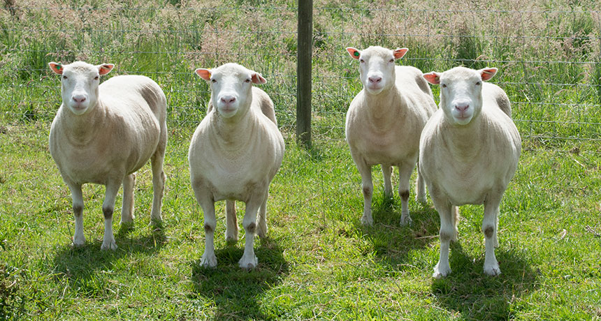
Clones don’t age prematurely, new research on Dolly the Sheep’s sisters suggests.
Researchers and animal welfare activists have been concerned that cloning, or somatic cell nuclear transfer, could cause health problems in cloned animals. Instead, a study of 13 cloned sheep found no signs of early aging or other health problems, researchers report July 26 in Nature Communications.
“These animals were remarkably healthy and fall within the normal range that we’d expect in animals of this age,” said developmental biologist Kevin Sinclair of the University of Nottingham in Leicestershire, England. Sinclair spoke July 25 during a news conference at the EuroScience Open Forum in Manchester, England. The cloning technique places the DNA-containing nucleus of an adult cell into an egg where the DNA is reprogrammed to an embryonic state. Dolly the Sheep, born in 1996, was the first mammal ever cloned. Since then, researchers have cloned a wide variety of animals. The technique doesn’t always work and many potential clones die before birth or shortly after. Surviving animals might have problems because of incomplete reprogramming of the DNA.
Dolly herself gave rise to the idea that clones age fast. Compared with other animals her age, Dolly had shorter telomeres, the caps that protect the ends of chromosomes from unraveling. Short telomeres have been associated with aging. Plus, Dolly had severe arthritis. She died at age 6, although not of old age. Dolly and other sheep in her flock were infected with a virus that killed them (SN: 3/1/03, p. 141).
Her untimely death, arthritis and short telomeres “were mushed together in people’s perception,” leading to the idea that clones age prematurely, said Katrin Hinrichs, a reproductive physiologist at Texas A&M University College of Veterinary Medicine and Biomedical Sciences in College Station. Hinrichs and other researchers not involved in the study hope the new report corrects the record on cloning and aging. “Now we have a reference to say what is and what is not a result of cloning,” she says.
How fast animals age varies, even among nonclones, says reproductive biologist Mark Westhusin, also of Texas A&M. Westhusin was on the team that produced cc (short for Carbon Copy), the first cloned cat (SN: 3/23/02, p. 189). She is now 15 and doing fine, says Westhusin. “This is a nice paper to confirm in a more formal scientific setting what most people involved with cloning have believed for a long time,” he says. Some studies have even hinted that clones may live longer than conventionally bred animals (SN: 4/29/00, p. 279).
In the study, Sinclair and colleagues examined 13 cloned sheep from 7 to 9 years old (roughly equivalent to people in their 50s to 70s). Four of the sheep — Debbie, Denise, Dianna and Daisy — were cloned in 2007 from the same mammary gland tissue that produced Dolly. “We had four almost identical sisters to Dolly and thought this would be a great chance to revisit this,” Sinclair said. He and colleagues compared the Dolly the Sheep sisters and nine clones of other sheep with 5- to 6-year-old sheep bred by traditional means. Cloned sheep had normal blood sugar, insulin levels and blood pressure. A few had mild arthritis. One of Dolly’s sister clones had moderate arthritis. The researchers have not yet measured the clones’ telomeres.
Sheep in this study were cloned with modifications to the original technique that may have produced a better outcome. But Dolly’s problems didn’t necessarily stem from being a clone. She may have developed arthritis as a result of trauma to her joints. It’s also not clear whether her short telomeres were really an indicator of premature aging. Certainly her death had nothing to do with being a clone; noncloned animals in her flock also died, researchers say. Overall, Sinclair said, “perhaps Dolly was a little less lucky.”
Cloning today is done mostly in South America and Asia, and infrequently in the United States, says Hinrichs. Polo ponies and cattle are among the most-cloned animals. “Cloning is so costly and inefficient that your animal has to be very special for a cloning to be worth it,” she says. As a result, most cloned animals are prized breeding stock or performance animals. Some animals that are genetically resistant to diseases are also cloned for veterinary and medical research.
When the brain runs low on oxygen, red blood cells sense the deficit and hurl themselves through capillaries to deliver their cargo. That reaction, described online August 4 in Neuron, suggests that red blood cells can both detect and remedy low oxygen.
When researchers stimulated the feet of mice, nerve cells fired off signals in the corresponding part of the brain, depleting that area’s oxygen. Red blood cells in capillaries picked up their speed in response. And in artificial capillaries, the lower the oxygen, the faster the red blood cells moved, Jiandi Wan of the Rochester Institute of Technology in New York and colleagues found. That swiftness was caused by the cells becoming more flexible, a bendiness that let them squeeze through narrow capillaries faster. When researchers stiffened red blood cells with a chemical, the effect of low oxygen on speed disappeared.
The results reinforce the complex and important role of blood in the brain. The findings might ultimately be relevant for disorders in which the link between neural activity and blood flow is damaged, including Alzheimer’s disease, says study coauthor Maiken Nedergaard of the University of Rochester Medical Center.