Apple announced on Tuesday a plan to open a new research and development (R&D) center in Shenzhen city, South China's Guangdong Province while upgrade its Shanghai R&D center to support product manufacturing.
The new move came as the company just announced to open a new store in downtown Shanghai, underscoring the US tech giant's confidence in the Chinese market.
Later in 2024, Apple will open a new R&D center in Shenzhen, which is expected to provide strong support for the company's staff in the whole region while deepening cooperation with its local suppliers. The new center will strengthen Apple's capability in the testing and research of products including the iPhone, iPad, and Vision Pro, the company said in a statement appeared on its China website.
It also plans to upgrade its Shanghai R&D center to provide support for the reliability, quality and material analysis of its products.
"In Apple, we are proud that we can deepen our footprint in China and expand our world-class facilities here," Ge Yue, Apple's vice president and managing director of Greater China region, was quoted as saying.
Ge said that the new investment will further implement the company's commitment in the market and provide world-class products for Apple users.
According to Apple, it has invested more than 1 billion yuan in its advanced application R&D centers, and the investment volume will continue to increase along with the opening of the new facility in Shenzhen.
Recently, Apple has stepped up operations in China although the US government continues to seek "decoupling" with China with intensified sanctions, baseless accusations and repeated provocations against Chinese companies.
Apple is going to add a new store in downtown Shanghai on March 21, which is reportedly the highest-standard Apple store in the Chinese mainland.
Meanwhile, Apple faces fierce competition from Chinese local smartphone brands including Huawei, Xiaomi and OPPO.
Apple iPhones struggled in the Chinese mainland market in the first six weeks this year, with sales plunging by 24 percent year-on-year, a report by market research organization Counterpoint Research noted on March 5.
Southwest China's Xizang Autonomous Region recorded a GDP growth rate of 9.5 percent last year, the highest in the country. Riding on the momentum, the region is expected to sustain its robust economic recovery in 2024, boosted by the development potential in specialty industries such as highland barley and yaks.It will also step up basic infrastructure construction as well as local tourism, a Chinese national political advisor told the Global Times during the two sessions.
Duoji Cizhu, a member of the National Committee of the Chinese People's Political Consultative Conference (CPPCC) and the president of the Xizang Federation of Industry and Commerce, said that Xizang enjoys a unique geographic advantage as it is an important passage connecting China with South Asia. The region could consider further high-level opening-up to facilitate the development of inbound and outbound tourism.
Last year, travel agencies in Xizang organized 23 outbound tourists group to Nepal, according to a report by media outlet chinanews.com.
Some deputies and political advisors also suggested Xizang should expand border trade with neighboring countries such as Nepal, which is Xizang region's largest trading partner and export market. Xizang and Nepal have inaugurated the jointly building of a China-Nepal industrial park in Nepal in 2019, which is also a key bilateral cooperation project under the Belt and Road Initiative (BRI). Xizang has also set up a border economic cooperation zone in Gyirong county.
In 2023, Xizang's foreign trade more than doubled to 10.98 billion yuan, local customs data showed. Its GDP expanded 9.5 percent last year, significantly higher than the national average of 5.2 percent. All those key indicators speak volume for the local economy vitality and a potential in development prospect.
"Xizang's basic infrastructure remains relatively undeveloped, so it is hoped that infrastructure building will gear up in 2024," Cizhu said.
Duan Xiangdong, a member of the CPPCC National Committee and the chairman of Aluminum Corp of China, said in addressing a proposal to the ongoing two sessions that the region, and in particular Ali prefecture, "a key area in China's opening-up to South Asia, which is also rich in natural and mineral resources," should double down on basic infrastructure investment, so that it is able to translate its resource advantages into economic dividends.
"The installed power grid capacity in Ali cannot meet rising electricity demand for social and economic development. Transportation in Ali is limited to highways, with no rail access, which is not cost-effective," Duan pointed out. He stressed that the development of the border regions is also conducive to ensuring national security.
Cizhu also serves as the president of Xizang business chamber. He envisioned that the private sector would play a more pivotal role in the development of overall local economy in 2024.
"Private enterprises in Xizang are set up by firms across the country, and they, while contributing to the improvements of people's livelihoods, also carry great significance in strengthening ethnic unity and the consolidation of border defense," he noted.
The work report of the Standing Committee of the 14th National People's Congress (NPC), China's top legislature, on Friday pledged to accelerate the formation of a law aimed at promoting the development of the private sector, sending a strong signal on policymakers' commitment to make continuous improvements in the business environment and boosting the high-quality development of the private sector for Chinese modernization.
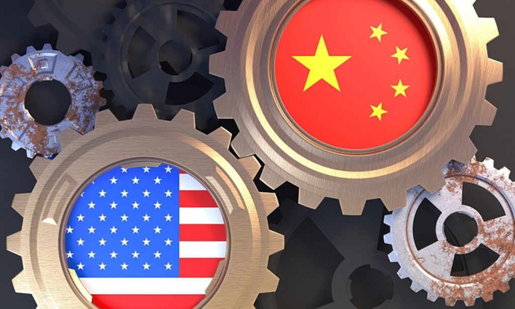
China's exports to the US reached 522 billion yuan ($72.52 billion) in the first two months of 2024, up 8.1 percent year-on-year, which analysts said reflects the strong competitiveness of Chinese products despite Washington's protectionism.
According to data released by the General Administration of Customs (GAC) on Thursday, the US remained China's third largest trade partner in January and February, with bilateral trade up 3.7 percent on a yearly basis to reaching 707.7 billion yuan.
China's imports from the US fell 7 percent to reach 185.7 billion yuan.
China's trade surplus with the US hit 336.3 billion yuan during the period, up 18.8 percent year-on-year, GAC data showed.
"The remarkable growth in China's export to the US during a slack season is uplifting, underscoring the important trade relations between the two countries," Zhou Mi, a senior research fellow at the Chinese Academy of International Trade and Economic Cooperation, told the Global Times on Thursday.
Although China and the US have engaged in dialogues in a variety of fields following the meeting between the two heads of state in San Francisco in November, they failed to reach consensus in many aspects. The US needs to send more cooperation signals to the market, Zhou said.
Dismissing Western media hyping up that Mexico replaced China as the top exporter to the US in 2023, Zhou said that Chinese goods are internationally competitive, and without Washington's political interference, there is still possibility that China and the US will continue to be each other's most important trade partners.
He said there is a great potential for China to export machinery products, electric vehicles and charging equipment to the US. While the US aims to revitalize its domestic manufacturing sector, China also has advantages in exporting intermediate products and components.
In response to the US investigation into Chinese made vehicles that use "connected" car technology, Zhou said the US' protectionist moves bring harms to market stability and global industrial and supply chains.
"If the US wants to reach some achievements in climate change and green development, it should fully give play to each country's advantages across the global market and reduce the trade barriers," he said.
Members of the Chinese People's Political Consultative Conference (CPPCC) National Committee have proposed building a science and technology innovation center in Northwest China's Xinjiang Uygur Autonomous Region oriented toward Central Asia, in a bid to promote the Belt and Road Initiative (BRI) and local development.
Chinese experts said that the center will facilitate technology cooperation and business trade between Xinjiang and Central Asian countries, further lifting personnel, technology and capital flows in the region.
The proposal was co-raised by three CPPCC National Committee members -- Liang Yong, Deng Mingjiang and Xiao Wenjiao -- aiming to foster Xinjiang's innovation-driven development mode, technological innovation capability and the new productive forces, the Global Times was told on Monday.
The proposal called for efforts in supporting Xinjiang to build multiple innovation bases with distinctive industries and advantages, and form new development modes in major cities of the autonomous region and BRI partner countries in Central Asia.
The proposal called for an enhanced coordination mechanism targeting technological support to Xinjiang, diversifying the supporting approaches guided by governments, and ensuring the deployment of talent and expertise.
Xinjiang has multiple advantages in academic research, industrial engineering, new energy and agriculture, which are highly complementary to the industries of Central Asian countries, said the proposal, noting that the innovation center will expand regional cross-border cooperation to stabilize the nation's energy supply and diplomatic relationships.
Liu Zongyi, director of the Center for South Asia Studies at the Shanghai Institutes for International Studies, told the Global Times on Monday that China will not only export self-developed technologies but also learn from some Central Asian countries' leading experience.
In addition, the proposal said the innovation center will amplify the advantages of local pilot free trade zones, balance the development in China's western and eastern regions, and attract international technology, talent and businesses.
"The innovation center will simplify Xinjiang's international cooperation with neighboring Central Asian countries, and will create mutually beneficial results for both sides," Liu noted.
China's cybersecurity technology ranks in the "top tier" globally, and in the realm of security and defense it can now stand on par with the US, Qi Xiangdong, chairman of Qi An Xin Technology Group, told the Global Times on Friday. However, there remains a gap between China and some developed countries such as the US in terms of investment in cybersecurity, Qi noted.
The rapid development of artificial intelligence (AI) brings new cybersecurity threats, and China should accelerate the integration of cybersecurity and AI technology to enhance its capability to deal with cybersecurity risks and uncertainties in cyberspace, Qi said.
Qi, who is also a member of the National Committee of the Chinese People's Political Consultative Conference (CPPCC), said during a group interview ahead of the opening of the two sessions this year that the development of AI technology has become a hot topic for discussion both domestically and internationally.
However, the security risks it brings have also raised concerns. Some experts estimate that over the next decade, the malicious use of AI technology will grow rapidly, posing serious threats to political security, cybersecurity and military security.
Regarding this hotly debated topic, Qi believes that there are three main types of new cybersecurity risks that would come along with AI technology.
First, AI itself exacerbates security threats, such as data breaches, fraudulent attacks, and security in social governance.
"Generative AI technologies represented by ChatGPT and Sora can quickly generate phishing emails and write malicious software and code, leading to an explosive growth in the number of attacks and frequent AI fraud incidents," he told the Global Times.
Moreover, criminals can use "deepfake" technology for face-swapping and voice manipulation, creating fake videos, so that "seeing may not necessarily mean believing" could become the norm.
The second type of security risk lies in the potential exacerbation of the "imbalance between offense and defense" in the field of cybersecurity, resulting in greater vulnerability to attacks, Qi noted.
AI significantly lowers the barrier to entry for cyberattacks, allowing ordinary individuals without coding or technical knowledge to become hackers, and thereby increasing the number of cyberattacks. Meanwhile, specialized hacker organizations can leverage AI tools to modify and upgrade their attacks.
The third type of risk is that AI exacerbates military threats, with the trend of AI weaponization becoming apparent, Qi said. He noted that AI can be used in lethal autonomous weapons like "killer robots," enabling autonomous identification of targets, remote automated operations, concealing the source of attacks, establishing advantages in confrontation, and connecting networks, decision-makers, and operators, making military actions more targeted, precise, and widespread.
In fact, an increasing number of countries are exploring the application of AI in the military domain.
Qi told the Global Times that the key source for AI is big data, so China must first of all solve data security issue to address the threat from AI, he said.
To tackle this challenge, a comprehensive approach is needed, involving not only "intelligence against intelligence" but also "coordinated development between humans and machines," Qi said.
Under the new circumstances, it is necessary to strengthen the promotion of technological innovation, encourage leading companies in various industries and cybersecurity companies to cooperate, integrate AI security technology into digital scenarios, and provide effective security protection, Qi noted.
At the same time, it is necessary to leverage AI capabilities to accelerate innovation in cybersecurity technology and security protection systems, in order to "run faster than AI technology," he said.
"Security is all about speed," Qi said, noting that effective security protection for new scenarios created by new technologies is essential for the continued promotion and application of technology; otherwise, technology applications will perish in their infancy.
AI can also be applied in the field of cybersecurity, Qi stressed. A security expert can handle 120,000 alerts in one year, and "our innovative Q-GPT security robot increases the efficiency of alert handling by 70 times compared to a human," Qi said.
This could help security experts save an average of 80 percent of their "screen-watching" time. It also allows them to use the time saved to engage in high-value tasks related to business and direct robots to handle more complex security incidents, Qi said.
Over the past year, international cyberspace competition has become more intense, with frequent cyberattacks. In response to this situation, China is strengthening the construction of its internal cybersecurity system, Qi said.
The cyber armies and intelligence agencies of some unfriendly countries will never cease their cyberattacks on China, just as we cannot rid the world of bacteria and viruses. Qi explained that the internal cybersecurity system is like the human immune system. "It can kill bacteria and viruses, or prevent these bacteria and viruses from affecting our health. That's its function," he said.
Although China's cybersecurity technology has reached the top tier globally, there is still a gap between the investment in cybersecurity by Chinese government departments and enterprises compared to that in the developed countries, Qi noted.
"The US 2024 fiscal year budget shows that the cybersecurity budget of civilian federal agencies accounts for approximately 16.4 percent of the IT budget, while in China, it's still around 3 percent, which is a huge gap that needs to be filled," Qi told the Global Times.
"Security is paramount, and insufficient investment will inevitably lead to insecurity," he said. According to the experience of developed countries, cybersecurity investment should account for more than 10 percent of the total IT investment to support digital business.
Chinese state-owned enterprises (SOEs) are giving full play to the main role of central SOEs in strengthening the development of artificial intelligence (AI) in response to the central government's call, the Global Times learned from companies and experts.
The move reflects the government's and industry players' determination to promote the advance of AI technologies in order to achieve an industrial transformation and upgrading, experts said.
In a recent statement sent to the Global Times, China Telecom, one of the major telecommunications operators of China, said the company gives full play to the main role of central SOEs and reinforces its strength in driving technological innovations.
The company has led the way after it released the 100 billion parameter Xingchen large language model in 2023, with more than 10,000 daily active users. The operator said it had made Xingchen open source at the end of January, a move that will allow for easier and broader collaboration.
"By doing so, we will broadly empower more users to engage in AI advances, injecting vitality into the AI industry," the company told the Global Times, adding that it has served more than 1 million users nationwide.
China Mobile, another telecommunications operator, is building Asia's largest intelligent computing center, which is scheduled to open this year, according to media reports.
The Chinese government has ramped up the promotion of AI development among SOEs. On Monday, the State-owned Assets Supervision and Administration Commission of the State Council (SASAC) held a meeting on promoting the reform of SOEs, stressing the importance of pushing forward the transformation and upgrading of central SOEs through technological empowerment, including AI.
The SASAC held a meeting on February 19 calling on central SOEs to accelerate the layout and development of the AI industry, actively promote industrial renewal and achieve better growth.
SASAC Chairman Zhang Yuzhuo emphasized at the meeting the need to promote central SOEs to achieve better growth and play a greater role in the field of AI.
The meeting was attended by representatives of SOEs in various industries, including telecommunication and information, manufacturing, transportation and energy, as well as high-tech firms such as iFLYTEK, according to media reports.
The SASAC also vowed to accelerate the construction of a new batch of intelligent computing centers and better leverage the role of the platform for collaborative innovation among SOEs.
Market analysts said that recent intensified efforts by the SASAC and enterprises underscored the country's determination and resolve to promote advances in AI technologies among SOEs to achieve transformation and upgrading, enhance competitiveness, and achieve other goals.
Li Jin, chief researcher at the China Enterprise Research Institute in Beijing, told the Global Times on Wednesday that the meetings can be seen as the "first shot fired" in AI deployment, and it is likely to propel a new wave of industrialization for SOEs.
China has its own advantages in the field of AI, Li said, noting that with strong policy support, a huge population, strong data collection and capabilities and innovation, China's AI development will surely reach the world's advanced level in the next decade.
According to data released by the SASAC, SOEs completed 2.18 trillion yuan ($302.8 billion) of investment in strategic emerging industries in 2023, up 32.1 percent year-on-year.
"AI is growing rapidly, and central SOEs should become key players on the 'national team,' in embracing AI technology, in the face of a new round of industrial revolution," said Li.
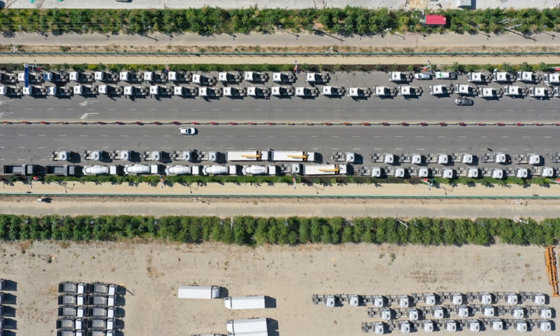
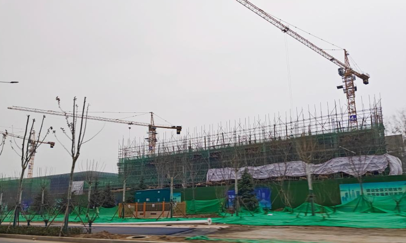
Many areas across China have commenced construction of mega projects since the Chinese Lunar New Year holidays, including industrial upgrading, infrastructure and energy projects, in a boost for the ongoing economic recovery, according to media reports on Monday.
On Monday, authorities in East China's Shandong Province held a meeting to promote the construction of major projects, with construction work for more than 1,000 projects started, China Media Group reported.
The projects involve a total investment of about 1.21 trillion yuan ($167.84 billion), according to local media reports in Shandong. Among the projects, 660 were for industrial upgrades and 156 for transport and other infrastructure. In terms of funding, 600 projects were invested in by private enterprises and 338 were carried out by state-owned enterprises.
Shandong is not alone in expediting major investment projects, as work resumed after the Chinese Lunar New Year holidays. In East China's Zhejiang Province, construction work has also commenced on 333 projects with a total investment of 977 billion yuan. In Beijing, 160 projects with a total investment of 247.8 billion yuan are expected to be launched in the first quarter of 2024.
The accelerated implementation of major projects across the country is expected to offer a great boost for investment, which remains a major economic driver, in the first quarter of 2024. This in turn will help support the economic recovery.
Relatively slow growth in investment has weighted on China's economic recovery. In 2023, total fixed-asset investment only grew by 3 percent year-on-year, compared to 5.1 percent growth in 2022. Some economists have forecast that fixed-asset investment could grow around 5 percent in 2024.
Private investment is also vital for driving overall investment, and Chinese localities have stepped up support for private businesses, especially since the Chinese Lunar New Year holidays.
Shandong has taken various measures to support the resumption of work at private businesses following the Chinese Lunar New Year holidays. For example, on Monday, State Grid Yantai Power Supply Co, the local branch of the state-owned power giant, dispatched a special working group to a local firm, Laizhou Sanli Auto Parts Co, which exports auto parts to Europe, North America and many other regions, to help the firm with issues in using electricity.
The China Chamber of Commerce to the EU (CCCEU) on Friday launched a financial working group and held a forum on cross-border yuan payment and trade cooperation, aiming to strengthen China-EU financial collaboration, even as businesses seek to navigate challenges posed by increasing trade protectionism in the EU.
The new working group, following the digital and green working groups, represents the CCCEU's third working group and its first outside its Brussels headquarters. It underscores the Chamber's dedication to enhancing the presence of Chinese enterprises in the EU market, striving for mutual benefits while acknowledging the diversity across the region.
The establishment of the financial working group aims to foster financial cooperation between the EU and China as well as to promote the internationalization of the yuan. This initiative is part of the CCCEU's efforts to implement the outcomes of the 10th China-EU High-level Economic and Trade Dialogue, Sun Yanhong, a senior research fellow at the Institute of European Studies under the Chinese Academy of Social Sciences, told the Global Times on Saturday.
As Chinese enterprises increase their investment in Europe and amid the increasingly complex China-EU trade situation, this move by the CCCEU will contribute to the internationalization of the yuan, facilitating trade and investment for Chinese companies, said Sun.
Trade protectionism has emerged as a significant barrier to the longstanding trade partnership between China and the EU. Recent actions, including an antitrust investigation into Chinese train-maker CRRC Qingdao Sifang Locomotive by EU regulators, illustrate the EU's shift towards more protective measures.
The EU and China are each other's major trade partners, with China's total imports and exports of goods with the EU reaching 5.5 trillion yuan in 2023, a decrease of 1.9 percent compared to the previous year, according to the General Administration of Customs.
But the EU's increasing trade protectionist measures are prompting concerns among businesses about the negative impact on the close trade ties between Europe and China.
In response to Europe's trade protectionist measures against China, Ola Kaellenius, CEO of German auto giant Mercedes-Benz, criticized the approach, stating that any move by the EU to increase protectionism against China would be a destructive move for an economic region like Europe, according to Reuters.
Trade protectionist actions, such as the investigations against Chinese electric vehicles and CRRC, not only harm the development of Chinese companies in the EU but also cause European corporations like Mercedes-Benz to worry about potential counter measures from China, Sun said.
With the EU implementing more trade protectionist measures and export control policies, it could lead to significant losses for multinational corporations, with ASML from the Netherlands being a prime example, Sun stated.
In January, the Dutch chipmaking equipment producer ASML, warned that the US' export controls could impact its sales in China by 10-15 percent in 2024.
China and Thailand will sign a permanent mutual visa exemption agreement on Friday, said Thai Prime Minister Srettha Thavisin, as China's outbound tourism to Southeast Asia is expected to return to the "golden age."
Srettha was quoted by Thai media as saying that, by promoting implementation of the visa-free policy, Thailand, whose GDP is highly dependent on tourism, is about to see domestic tourism market flourish again following the return of Chinese tourists.
Srettha said on Wednesday in a keynote speech delivered at "Thailand 2024 The Great Challenges" that Thailand and China will sign a reciprocal visa exemption program this week. For Thailand's soft power, "the visa exemption program between the two countries is expected to upgrade the Thai passport's power to a higher level," he said.
Chinese Foreign Ministry spokesperson Wang Wenbin told a press conference earlier this month that the government departments responsible for the matter are in close communication on the specifics, after Srettha announced that Thailand and China will permanently exempt each other's citizens from visa requirements, starting from March.
The move will further enhance people-to-people exchanges and mutually exempt visas between China and Thailand serves the fundamental interests of both peoples, Wang said.
In September, Thailand implemented a five-month visa-free policy for Chinese tourists, which will continue until February 29, 2024. Chinese experts said the upcoming Spring Festival will see a new wave of people-to-people exchanges between the two countries as the expected visa exemption program will help tourism and economic and trade ties.
There are also signs that the tourism markets are expanding for both China and Thailand, and China's outbound tourism to Southeast Asia is expected to return to the poast "golden age".
"Between January 8-14, Thailand's inbound tourism performed 'better than expected,' mainly due to the smooth recovery the tourism markets in the region. Meanwhile, among the international tourists Thailand received during the period, Chinese tourists topped the list, increasing by 27.75 percent , compared to the number of 80,000 in the previous week," said Thai Tourism and Sports Minister Sudawan Wangsuphakijkosol.
Thailand beat the target of receiving 28 million foreign tourists in 2023, but the 1.2 trillion baht ($ 56.21 billion in yearly tourism revenue fell short of planned 2.38 trillion baht ($66.89 billion), the ministry said.
On online travel platforms, Thailand is most popular destinations for Chinese tourists traveling overseas during the Spring Festival holidays, according to a report sent by Qunar sent to the Global Times on Tuesday.
Tongcheng Travel said that the popular destinations for outbound travel during the coming holidays include Bangkok, Chiang Mai and Phuket.
The decision to sign an agreement on mutual visa exemption between China and Thailand has huge significance for both sides, especially for Thailand, a country which highly dependent on tourism to boost its economy. It will gain lots of benefits from being the largest tourist destination for Chinese tourists, Xu Liping, director of the Center for Southeast Asian Studies at the Chinese Academy of Social Sciences, told the Global Times on Thursday.
The mutual visa exemption will inject stronger momentum into the close bilateral relationship and accelerate people-to-people exchanges, expand economic cooperation, with a view to building a China-Thailand community with a shared future, said the expert.
China's annual Spring Festival travel rush, also known as chunyun, kicked off on Friday, and saw tens of millions of people rush to take trains, planes and other packed vehicles to reunite with their friends and families on the first day. Red lanterns, Chinese knots and bamboo dragons were hung up at train stations and airports, and passengers with luggage and gifts formed a vivid display of a robust and vibrant Chinese economy.
As the country braces for the largest annual human migration on the planet with a record 9 billion domestic trips expected, analysts projected a fresh consumption boom for the economy, which will not only give a boost to the world's second-largest economy, but also stimulate the confidence of enterprises to step up investment and innovation.
Dismissing the US-led Western bad-mouthing of the economy, they expressed full confidence in the Chinese economy's prospects, saying the country's institutional advantage in swiftly and effectively turning top policy blueprints into real actions will ensure the continuous stable upswing recovery of the economy in 2024, which is expected to grow by around 5 percent.
Fun and festive
China is expected to see more than 182 million passenger trips on Friday, the first day of its 40-day Spring Festival travel rush, according to a government estimate.
The hustle and bustle of chunyun was on vivid display at Beijing West Railway Station, one of the busiest train stations in the country. On a chilly morning, at around 7 am, the station was already crowded. Some passengers got off taxis before they even reached the drop-off point, and walked into the station pulling their luggage.
The noisy station was filled with vibrant colors and joyful laughter. Red lanterns, paper-cuts, Chinese knots and bamboo dragons could be seen at the station and on trains, immersing passengers in the fun and festive atmosphere of the upcoming Spring Festival.
"I couldn't wait to return to my hometown this Spring Festival and got up at 3 am this morning. When I arrived at the station, the festive decorations and cheers from the large crowd of passengers made me feel like the Spring Festival is already here," a 35-year-old worker named Li He told the Global Times on Friday.
On Friday alone, Beijing West Railway Station was estimated to have handled 143,000 passengers, and is expected to see a total of 4.92 million passengers, a spokesperson for the station told reporters on Friday morning. Operations were smooth with many workers guiding passengers quickly through security checkpoints and to their departure gates - a reflection of the improved capacity and services of China's ever-expanding modern transport network.
On Friday, China Eastern Airlines debuted all four of its domestically-produced C919 large passenger jets, carrying 111 passengers from Shanghai to Chengdu, Southwest China's Sichuan Province by noon. In the country's civil aviation sector, a total of 2 million passenger trips are expected to be handled on Friday, up 94 percent from the same day in 2023 and a 15 percent rise from that in 2019.
At Beijing Daxing International Airport, a passenger named Chen Peng told the Global times that he is taking his family on a trip to Northwest China's Xinjiang Uygur Autonomous Region.
"It's the first time that we are taking my 6-year-old daughter to such a far region. In 2023, I got promoted with a higher income, and I hope to find a better life for my family and let my daughter see that our country is becoming better and better," Chen said. Boosting confidence
Extending the strong momentum during Spring Festival, the country's consumption sector - be it in cities or villages, online and offline, and in green spending or the silver economy - will continue to rebound robustly, with retail sales expected to exceed 50 trillion yuan ($7.04 trillion) in 2024, said Wei Jianguo, former Chinese vice minister of commerce and executive deputy director of the China Center for International Economic Exchanges.
This will not only play an important role in driving the Chinese economy but also in stimulating the confidence of enterprises, especially private companies, in stepping up investment and innovation, Wei told the Global Times.
"The year 2024 is the Year of the Dragon in the Chinese calendar. It is also the year that the Chinese economy will continue to keep an upward recovery trend, just like 'a dragon raising its head'," Wei said.
In response to the US-led Western campaign bad-mouthing and smearing China's economy after the release of the two economies' GDP data, analysts said the rapid economic growth of major economies contributes to global economic recovery and will help elevate the confidence of investors and consumers across the world. They expressed full confidence in the prospects of the Chinese economy, projecting that it will maintain around 5 percent growth in 2024.
Although the US' nominal GDP in 2023 of 6.3 percent is higher than China's 5.2 percent, the US' real GDP is 2.5 percent, data showed. "Part of the US' high nominal GDP growth rate is boosted by inflation and appreciation of the US dollar. Once these factors ease, China's GDP growth rate will be faster than that of the US," He Weiwen, senior fellow from the Center for China and Globalization, told the Global Times on Friday.
In addition, even the US' nominal GDP growth rate has shown a declining trend over the past several years, down from 9.2 percent in 2022 and 10.1 percent in 2021, which shows that the US economy is not as strong as some US politicians have touted, He Weiwen said.
The first and foremost challenge for the US economy is repeated standoffs due to its debt ceiling. If US debt continues to rise, it will definitely cause a US dollar crisis, Li Daokui, director of Tsinghua University's Academic Center for Chinese Economic Practice and Thinking, said at the World Finance Forum on January 20.
He Weiwen said the Chinese economy will continue to maintain overall stable recovery in 2024. Compared with specific GDP goals, it's more important to stabilize the property sector, dissolve local government debt risks, nurture new growth points, boost the continuous consumption rebound and improve the external environment, he said.
He said China must be firmly committed to opening-up by actively conducting cooperation with all economies and introducing outstanding knowledge, technologies and resources to develop new productive forces.
On Monday, a meeting of the State Council pledged efforts to enhance the innovation and coordination of policy tools, consolidate and strengthen the trend of economic recovery, and promote the stable and healthy development of the capital market.
Following the key meeting, multiple government agencies including the People's Bank of China, National Financial Regulatory Administration and National Development and Reform Commission, China's top economic planner, announced a series of measures, with the cut of the reserve requirement ratio by 50 basis points starting from February 5 being an important move.