CNOOC reports discovery of huge gas field in South China Sea
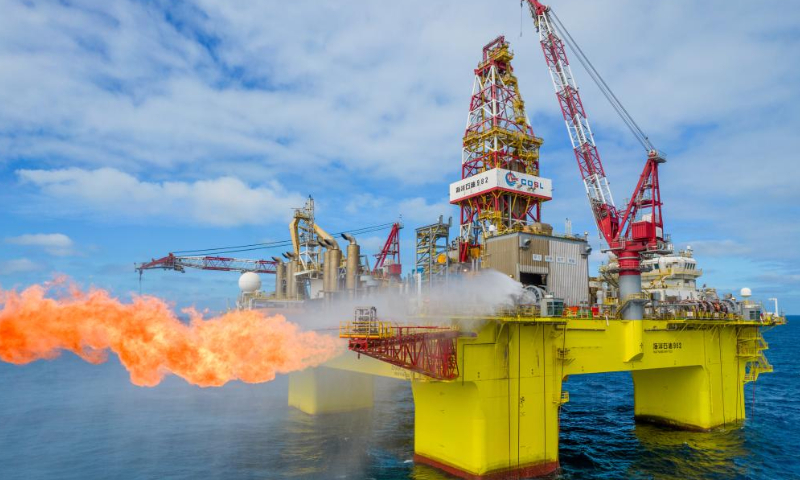
China National Offshore Oil Corporation (CNOOC) announced on Wednesday that the world's first ultra-deep water and ultra-shallow large gas field has been discovered near South China's Hainan Province and approved by national authorities.
The Lingshui 36-1 gas field, located in the South China Sea, has proven geological reserves of over 100 billion cubic meters of natural gas. The average water depth of the gas field is about 1,500 meters, and the average buried depth of the gas layer is 210 meters.
"The proven reserves indicate abundant future clean energy resources, which are crucial for China given its high demand for natural gas and petroleum. With import dependency at around 40 percent and natural gas being cleaner than coal, this discovery is vital for meeting demand, transforming the energy structure, and ensuring energy security," Lin Boqiang, director of the China Center for Energy Economics Research at Xiamen University, told the Global Times on Wednesday.
"To ensure offshore drilling safety, the industry usually avoids shallow gas zones in well design. This time, we took on the challenge directly," said a representative from a branch of CNOOC.
The discovery of Lingshui 36-1 offers valuable insights for similar offshore exploration globally. It also enhances China's deep-water exploration technology and completes the final piece of a plan to establish a 1 trillion cubic meter gas zone in the South China Sea, a deputy chief exploration engineer at CNOOC was quoted as saying in a Xinhua report on Wednesday.
In 2018, CNOOC initiated a plan to establish the "South China Sea Trillion Cubic Meter Gas Zone" by 2025. So far, it has confirmed over 1 trillion cubic meters of natural gas reserves in the Yinggehai, Qiongdongnan, and Pearl River Mouth basins, making the plan a reality.
In the first half of the year, China's natural gas production rose 4.4 percent to 123.5 billion cubic meters, while imports increased 14.8 percent to 90.2 billion cubic meters. For 2024, consumption is projected at 420-425 billion cubic meters, up 6.5-7.7 percent. Production is expected to reach 246 billion cubic meters, with a steady increase of over 10 billion cubic meters annually. Imports from the China-Russia East-Route are also set to grow, according to a report released by the National Energy Administration on July 23.